Technology
Technology
– PRF (Platelet Rich Fibrin)
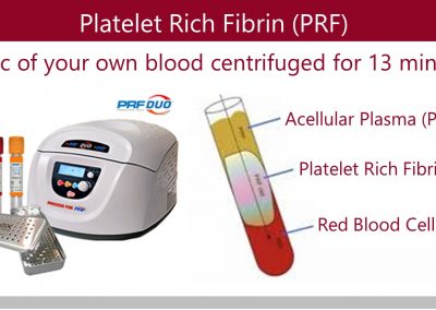
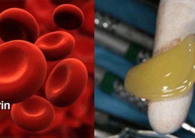
 Platelet Rich Fibrin is a by-product of blood that is exceptionally rich in platelets. PRF has long been used in hospitals to accelerate the body’s own healing process, but it is only fairly recently that advances in technology have allowed this same technique to be used in the periodontal office. A simplified chairside procedure results in the production of a fibrin membrane that is capable of stimulating the release of many important growth factors involved during wound healing processes that take place after surgery.
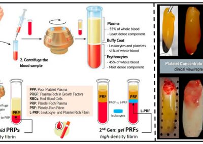
Platelet Rich Fibrin is derived from a small amount of a patient’s blood which is centrifuged and processed to separate red blood cells from platelets and leukocytes which form the basis of a complex fibrin matrix membrane. When a PRF matrix membrane is placed over a wound, the platelets are activated releasing several regenerative proteins with real therapeutic potential.
Essential regenerative proteins and growth factors include:
- PDGF – Platelet-Derived Growth Factor
- Healing Matrix Proteins
- Cytokines
- Transforming Growth Factor-B
- Insulin-like Growth Factor
- Vascular Endothelial (blood vessel lining) Growth Factor
- BMP – Bone morphogenetic protein
– DEKA Laser Assisted Surgery
 The CO2 Laser has been used in medicine for over 25 years and the DEKAÒ UltraSpeed CO2 Laser makes procedures virtually pain-free, speeds recovery, and improves periodontal surgical procedures in general. We utilize Laser technology to treat peri-implantitis (inflammation-bone loss) around implants and prevent implant failure.
All Lasers work by delivering concentrated light to where they are focused on. The DEKAÒ lasers cut through tissues in a manner that usually provides hemostasis (Lack of bleeding) and minimal post-operative discomfort. Often less anesthesia is required.
iCAT Cone Beam Digital Technology
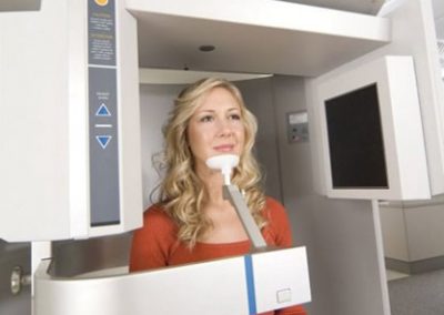
Dental cone beam computed tomography (CT) is a special type of x-ray equipment used when regular dental or facial x-rays are not sufficient. Your doctor may use this technology to produce three dimensional (3-D) images of your teeth, soft tissues, nerve pathways and bone in a single scan.

Dental cone beam imaging provides Dr. Derhalli with more accurate information and 10 times less radiation to the patient than the machines traditionally used in medicine. What this means to patients who have chosen to replace missing teeth, is that now Dr. Derhalli can examine, diagnose and plan treatment with a level of precision not possible in the past. This more exact approach using dental cone beam imaging means fewer complications, less invasive therapy, and quicker healing times for the patient.
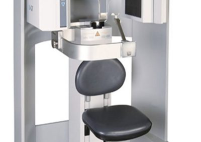
The iCAT Cone Beam Digital Technology produces more thorough three-dimensional views of all oral and maxillofacial structures. Patients remain seated in an “open environment scan,’ which increases comfort, and captures the natural orientation of anatomy. Once the data is captured, it’s transferred to a computer within minutes, and displayed on an intuitive 3-D mapping tool that allows doctors and technicians to easily format and select desired ‘slices’ for immediate viewing.
The New Dental Digital Implant Workflow
iTero™ Element Scanner
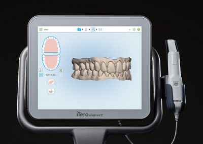
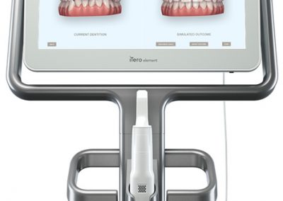
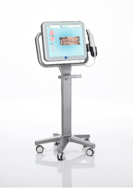
The new dental digital implant workflow begins with an accurate and fast scan that involves little to no patient preparation. The iTero Element Scanner is a state-of-the-art digital impression system that eliminates the need for messy putty in your mouth. The Element Scanner utilizes the latest advances in artificial intelligence (AI), is statistically more accurate than conventional impressions, can image dynamic occlusion, and can even reproduce realistic tissue colors.
Once the scan is obtained the scanning data is imported into a digital Computer Aided Design (CAD) software where real-time 3D modeling and manipulation can occur. This is often referred to as the modeling or design phase. Dental prosthetics (Implant Crowns) are manipulated in real-time 3D restorations, shaped, positioned, and finalized. This takes all the guesswork out of the process and guarantees a high degree of accuracy.
During the impression process, you can breathe or swallow as you normally would. You can even pause during the process if you need to sneeze or just want to ask a question. The scanner gives us a 3D model of your mouth that can be used for your dental services including Implant Crowns and Night Guards
BioMet Article
– 3-D Cone Beam CT Scan
The i-CAT cone beam imaging system provides 3-D technology with the capability of delivering a continuum of services from diagnosis to treatment. From less radiation for the patient to a typical scan time of only 8.5 seconds and reconstruction in under 30 seconds, the i-CAT imaging is quick, easy, and more thorough for the practice. We are able to virtually treatment plan each case utilizing in office scanning with three dimensional digital reconstruction of the patient’s anatomy. The 3-D anatomically accurate imaging allows me to create the most complete treatment plans benefiting patients with the most predictable surgical outcomes.
– Ostell Device (Implant Stability Evaluation)
Osstell ISQ is a complete diagnostics system used to determine dental implant stability. Accurate measurements of implant stability provide valuable diagnostic insight that helps ensure successful treatments. It provides me with accurate, consistent and reliable stability measures needed for making informed load decisions, avoid failure and give patients added quality assurance. Today, more patients ask for immediate loading of their implants.
The Osstell ISQ meter stimulates a SmartPeg™ mounted on the implant, by emitting magnetic pulses. These cause the SmartPeg to resonate with certain frequencies depending on the stability of the implant. The resonance is picked up by the Osstell ISQ meter. The SmartPegs have been calibrated in such a way that they all show comparable values for the same degree of stability, even when measuring implants from different systems.
– Advanced Surgical Software and Guided Surgery Techniques

Restoration Based Implant Planning
Anatomage supports both crown-down and implant-up treatment planning methods. This is possible by combining other types of clinical data with the CT scan, such as a laser scan of the stone models or an intra-oral scan of the dentition (digital impression). Additionally, virtual wax-ups can be incorporated as well. This allows me to plan precisely how the implant will be positioned with the future restoration (crown, bridge or denture) and occlusion of the patient.
– Digital X-rays (Digital Radiography)
One new dental technology involving dental X-rays is digital X-rays. They offer the advantage of an 80 percent reduction in radiation, no need for film or processing chemicals, production of a nearly instantaneously image, and the ability to use color contrast in the image. Digital radiography is a form of X-ray imaging, where digital X-ray sensors are used instead of traditional photographic film. Advantages include time efficiency through bypassing chemical processing and the ability to digitally transfer and enhance images. Also less radiation can be used to produce an image of similar contrast to conventional x-rays.
Instead of X-ray film, digital radiography uses a digital image capture device. This gives advantages of immediate image preview and availability; elimination of costly film processing steps; a wider dynamic range, which makes it more forgiving for over- and under-exposure; as well as the ability to apply special image processing techniques that enhance overall display of the image.

